

Melanoma is a type of skin cancer of very aggressive nature. If not diagnosed and treated in time, it can kill you in a few months. When caught early, however, melanoma is highly curable. Mole Mapping is an innovative method for early detection of melanoma. It allows us to identify subtle but suspicious changes in preexisting moles, as well as to detect new moles.
If you are considering Mole Mapping, here are some Frequently Asked Questions to get you started. If we don’t answer all your questions here, call us! And stay-tuned this month for more answers on Skin Cancer prevention, detection, and treatment during our Facebook Live Event with Board Certified Dermatologist, Dr. Ciro Martins.
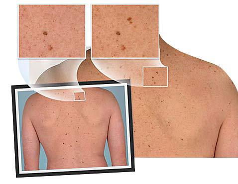
Shows Mole Mapping by Total Body Photography
Q: What is Mole Mapping?
A: Mole Mapping is a photographic documentation of your entire body surface done in a standardized fashion, using a high-resolution camera, therefore, providing a baseline set of images showing all the moles, their location, size, and what they look like, at the starting point, or baseline. These images are provided to you as an album of printed, large size photos and/or a disc with the set of digital images. The electronic image files are also downloaded into your EMR (Electronic Medical Records). These images will be your point of comparison for years. Every time you come back for a skin check, these images are used for comparison as needed. Whenever there are significant changes noticed on any particular mole, a biopsy will be done. If a new mole is found, and it was not there on the baseline photos, it will also be examined further and likely be biopsied if found to be suspicious.
Q: How does it work?
A: You schedule an appointment with our Medical Photographer, who will take a series of digital photos of your entire skin surface in standardized positions. The Dermatologist may indicate if any moles require more detailed photos. We store all digital images in your electronic medical records, so our dermatologists can review them for changes at your future visits.
Q: How often do I have to visit?
A: Mole Mapping requires a single appointment with the Medical Photographer and subsequent visits with our dermatologists. The frequency of your subsequent visits with the dermatologist depends on your skin cancer history and level of risk.
Q: Why are unusual moles photographed instead of removed?
A: Many moles we photograph are not necessarily suspicious for melanoma – however, they may have unusual features that defy some of the criteria that are established for typical moles. Mole mapping allows us to reduce unnecessary removals of benign moles and prevent melanoma by documenting change over time. If we detect a mole that is suspicious for melanoma, we will notify you immediately and arrange for its removal.
Q: Am I a good candidate for Mole Mapping?
A: We recommend Mole Mapping for those who are at higher risk of developing melanoma. High-risk includes, but is not limited to anyone with over 50 moles, anyone with a history of more than 2 blistering sunburns in childhood, anyone with a history of chronic tanning bed use and multiple moles, individuals with many atypical moles and also individuals with a personal or family history of melanoma.
Q: What if I’m too embarrassed?
A: At Belcara Health, we strive to make every patient feel as comfortable as possible. We have both female and male practitioners and a chaperone is available for the examination.
Q: Will I get to keep the photos?
A: A complete set of photographs and a CD will be sent to you and your physician (upon request).
Mole mapping is a very effective tool for the early detection of cancerous moles. If you are concerned about developing melanoma, contact us today to schedule an appointment: (410) 296-0414.